Publications

  • Diagram showing AI-mediated reading of fish freshness through microbial activity detection. It illustrates a process with three steps: hydration, spoilage sensing, and spoilage detection. The image includes a fish with a sensor attached, a smartphone scanning the fish, and a comparison between fresh and spoiled fish based on microbial presence.

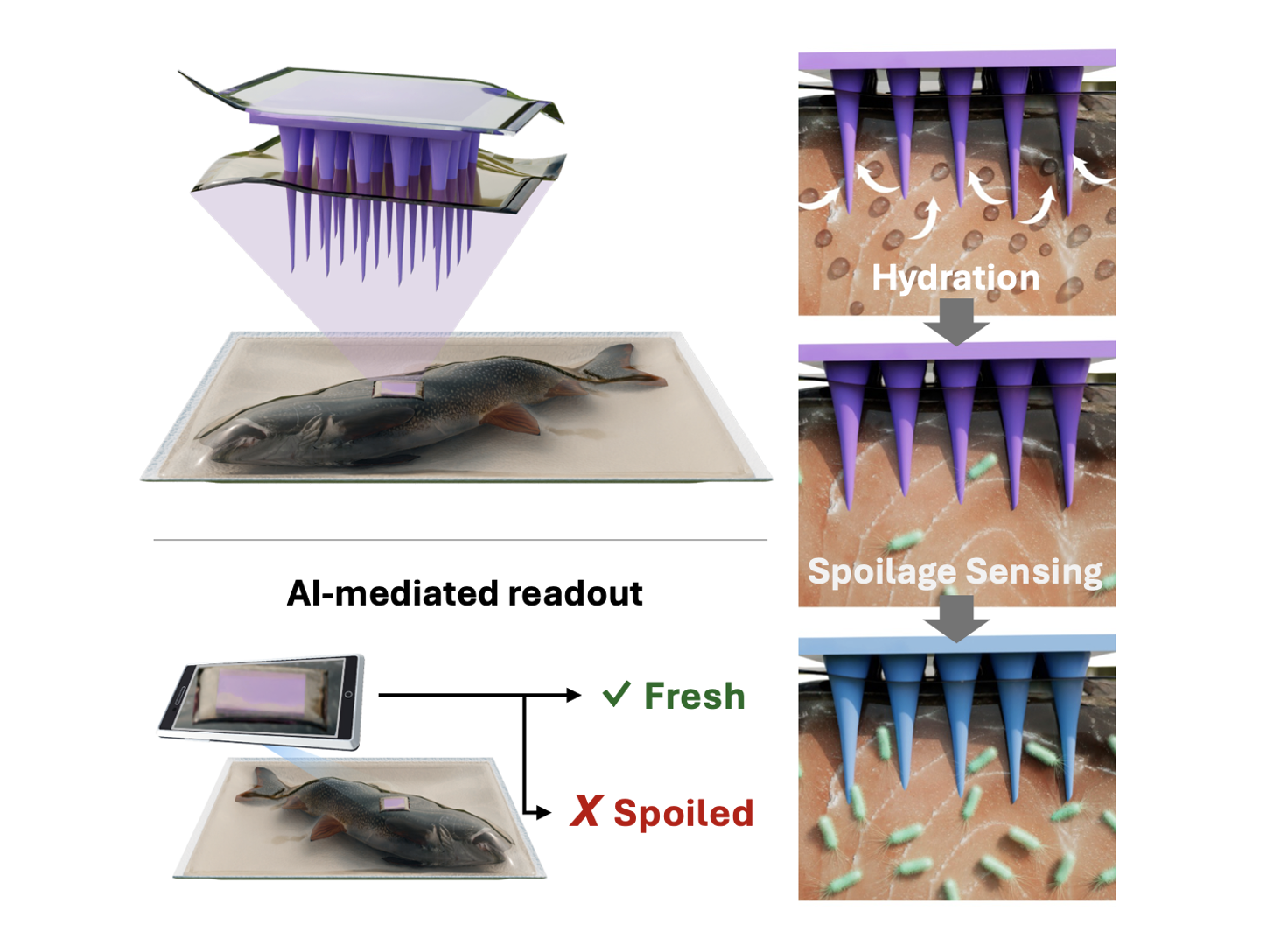
    Food-activated Microneedle Sensor for Real-time, Colorimetric Spoilage Monitoring of Pre-packaged Food

    Advanced Science, 2025

  • Diagram depicting the process of using bacteriophage nanomedicine for food safety. The top section shows how phages are combined with nanomaterials to target pathogens on food surfaces, leading to bacterial lysis and release of progeny phages. The bottom section compares decontamination methods: phage-loaded nanomaterials on raw beef versus surface phage delivery, showing the former achieves near 100% bacteria clearance according to a bar graph.

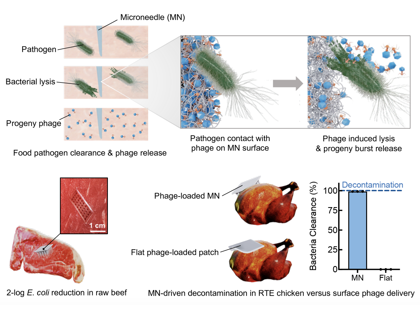
    Bacteriophage-Loaded Microneedle Patches for Targeted and Minimally Disruptive Foodborne Pathogen Decontamination

    Science Advances, 2025

  • Diagram illustrating different types of micro and nano-bots with physical, chemical, and biological propulsion methods. Top row shows magnetic, optical, acoustic, and electrical propulsion. Middle row depicts chemical propulsion with self-electrophoretic, self-diffusiophoretic, and bubble propulsion mechanisms involving platinum, silicon dioxide, and magnesium cores. Bottom row illustrates biological propulsion using spermatozoa, bacteria-based, algae-based, and macrophage-based bots.

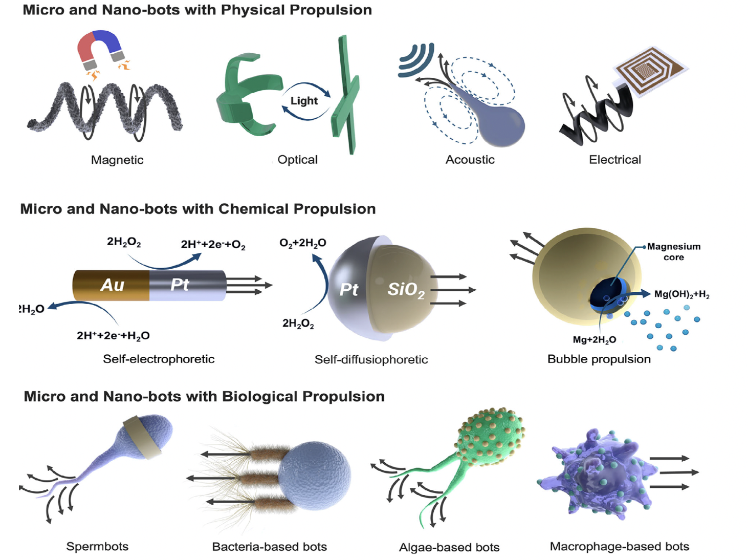
    Micro‐and Nano‐Bots for Infection Control

    Advanced Materials, 2025

  • Diagram showing the process of biological sensing. Top sections depict stimuli like blood flow, temperature, and strain; molecules such as gases, sugars, and metabolites; and targets like nucleic acids and amino acids. The middle section illustrates a sensor detecting sweat, tears, saliva, and interstitial fluid using light source, optical response, and detector components. The bottom sections detail aspects of the sensor's design including optical design, power management, data transmission, resilience, and wearability.

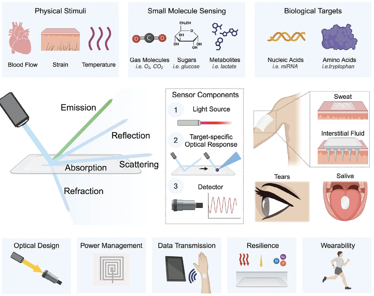
    Advances and Applications of Wearable Photonic Sensors

    Advanced Materials, 2025

  • Diagram showing medical and technological components, including antennas, tissue types, nanoparticles, electrodes, microfluidic layers, and devices used for monitoring and diagnosing conditions such as breast tissue, osteoporosis, and basal body temperature.

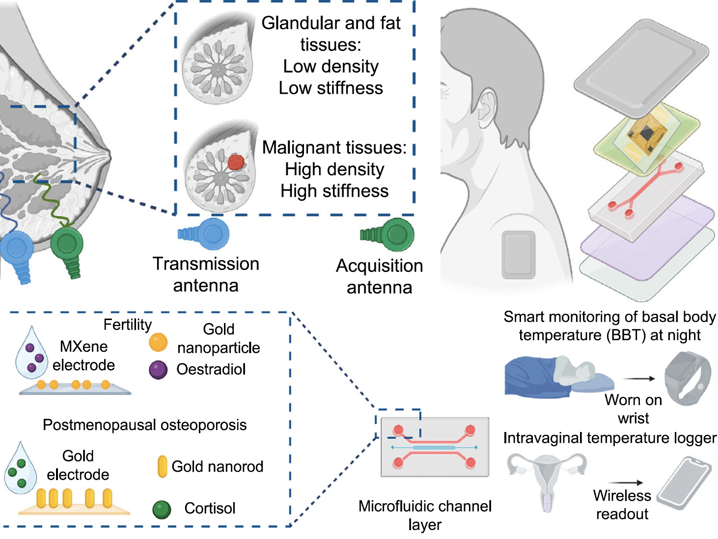
    Advances in Biomonitoring Technologies for Women’s Health

    Nature Communications, 2025

  • Diagram showing a process of 3D bioprinting of a bioink for immunofluorescence analysis. It includes a micro-arrayer printer with two bioinks: Bioink #1 with BSA and CAb in PBS, and Bioink #2 with EDC in MES. Bioink #1 is deposited first containing BSA molecules, while Bioink #2 contains active carboxyl groups for chemical bonding. The printed structure is analyzed using immunofluorescence to detect cytokines, biotin, and IL-6 with specific antibodies, visualized with fluorescent imaging.

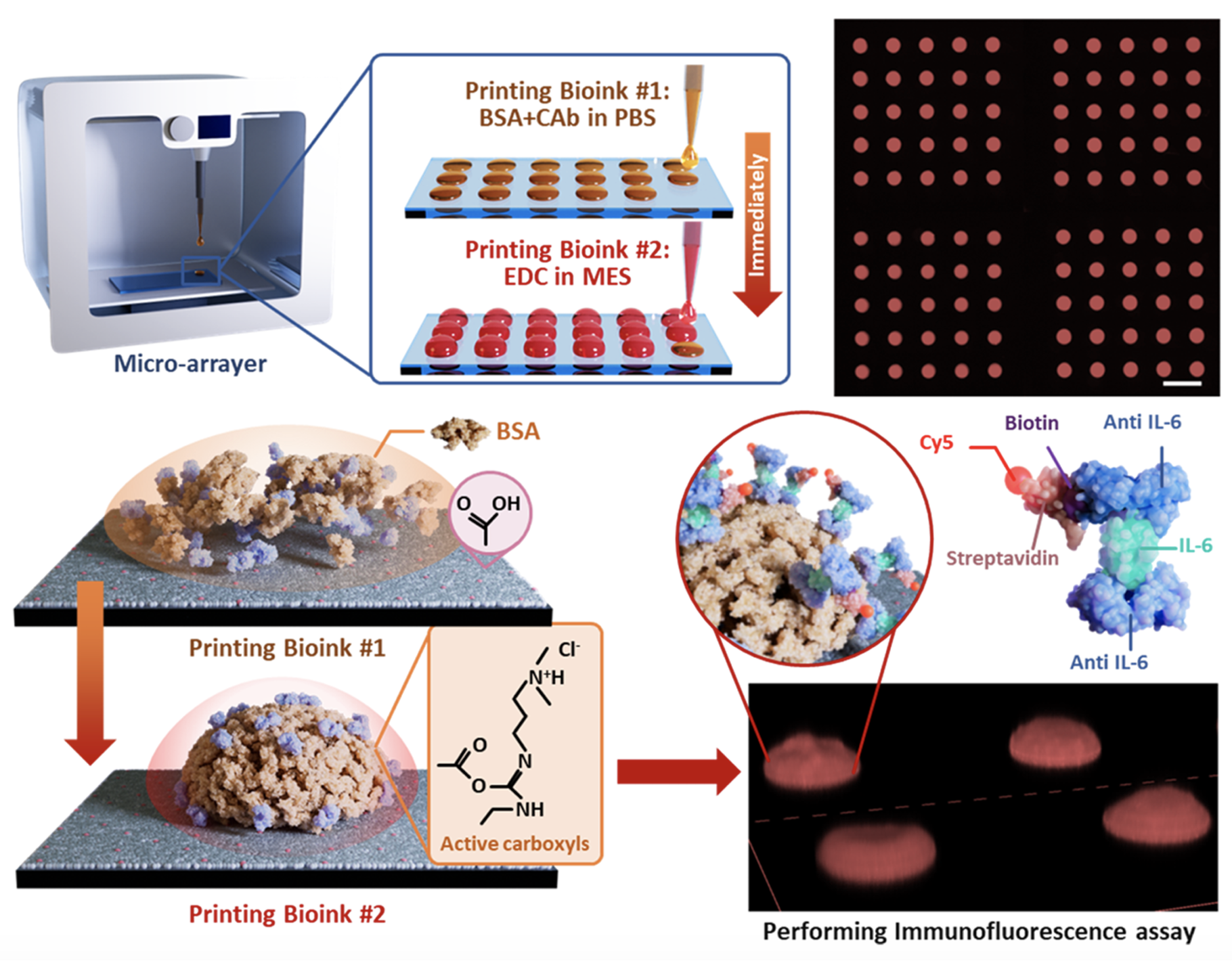
    Noncontact 3D Bioprinting of Proteinaceous Microarrays for Highly Sensitive Immunofluorescence Detection within Clinical Samples

    ACS Nano, 2024

  • Diagram illustrating the process of creating a honeycomb film using a PS solution in a humid chamber, water vapor condensation, solvent evaporation, and honeycomb formation, followed by the fabrication of phage microgels through adding phages, gelation, and peeling. Contains chemical reactions of phage cross-linking and the formation of ordered nanofibrous structures.

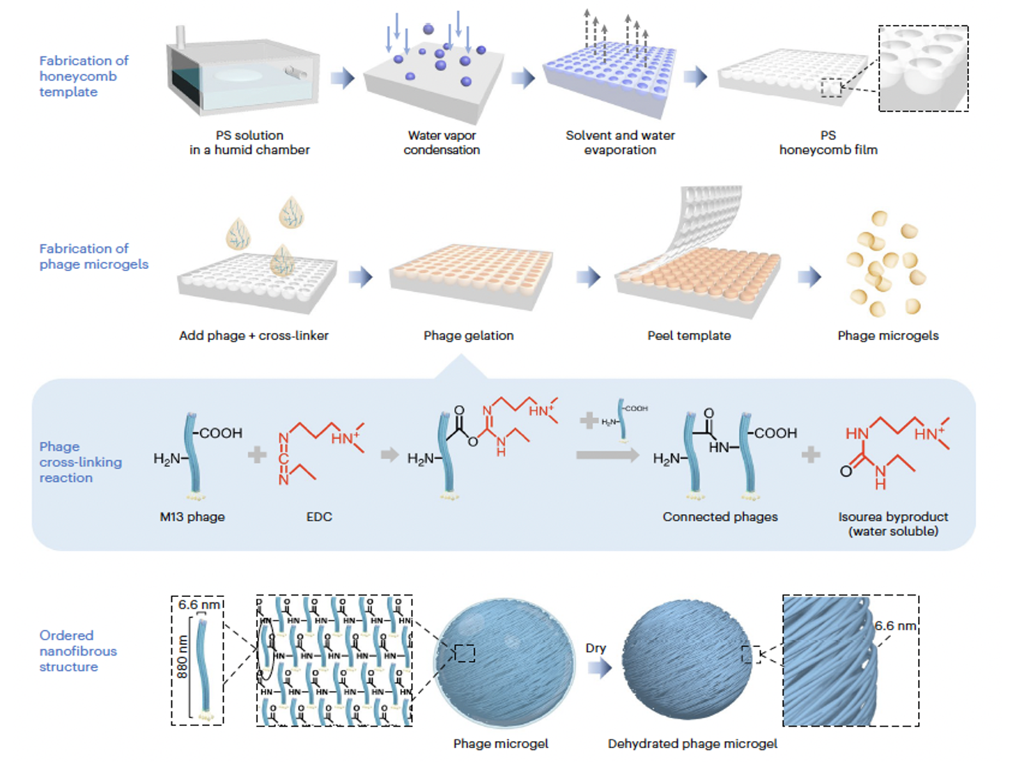
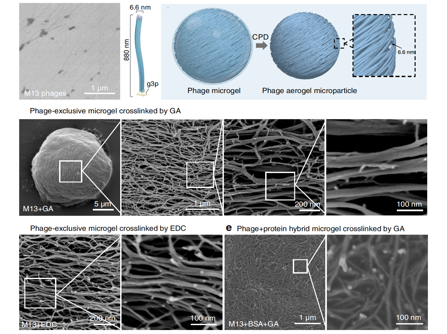
    High-throughput Fabrication of Antimicrobial Phage Microgels and Example Applications in Food Decontamination

    Nature Protocols, 2024

  • A collage of six close-up images of various flowers and floral structures, including a pink flower, a sunflower, a green bud, and abstract flower patterns.

    Virus‐Assembled Biofunctional Microarrays with Hierarchical 3D Nano‐Reticular Network

    Advanced Functional Materials, 2024

  • Diagram illustrating the process of targeted DNA cleavage using a DNAzyme complex attached to a polyacrylamide backbone. The complex includes a cleavage site, DNAzyme/substrate components, and an acrydite group. The diagram compares uncontained and contaminated scenarios with target proteins, oligonucleotide fragments, gold nanoparticles, and phages, showing DNA crosslinks. It depicts phage infection, DNA crosslinks, and subsequent target protein release, ultimately leading to DNAzyme cleavage and gold nanoparticle release.

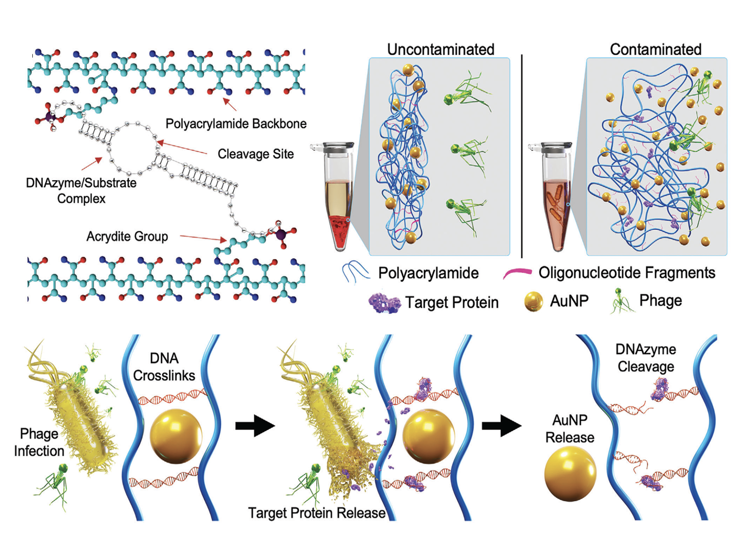
    Bacteriophage‐Activated DNAzyme Hydrogels Combined with Machine Learning Enable Point‐of‐Use Colorimetric Detection of E. coli

    Advanced Materials, 2024

  • Diagram showing fluorescence spectroscopy and detection methods. A graph plots fluorescence intensity versus wavelength, highlighting a specific wavelength around 400 nm with a check mark. A color wavelength scale ranges from blue to red, pointing to a piece of meat. Two laser diagrams indicate high signal-to-noise ratio and fluorescence detection at the meat sample.

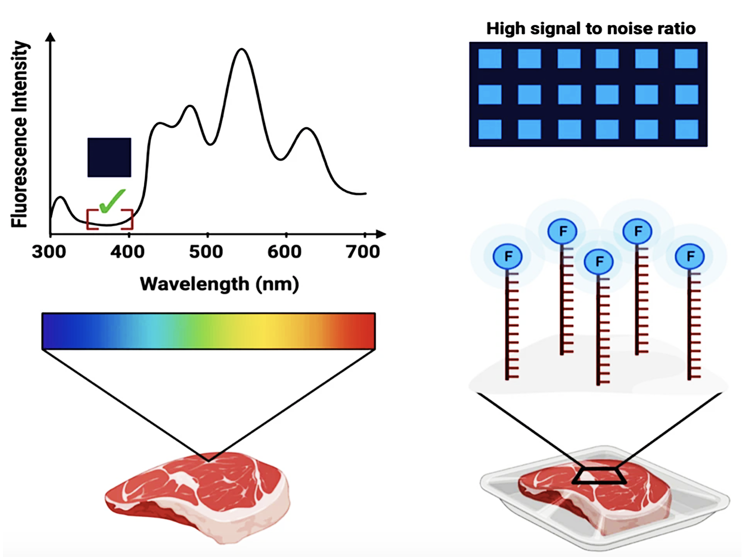
    Fluorescence Profiles of Contamination-prone Foods Applied towards Microcontact-printed in situ Functional Oligonucleotide Sensors

    Scientific Reports, 2024

  • Diagram showing bacterial detection using fluorescence imaging. It includes a piece of meat, a saturated membrane with an FNAP sensor, and a fluorescence imaging device. The diagram depicts bacteria labeled with CAR T cells, phages HER262 and T7, each with tail structures.

    Down to Business: Smart Food Packaging Commercialization

    Nature Reviews Bioengineering, 2024

  • Comparison of molecular structures after contamination and light exposure. Top row shows OmniKill molecules; left, after contamination, molecules are intact; right, after light exposure, molecules have been destroyed. Middle row shows Repel molecules; left, after contamination, molecules are intact; right, after light exposure, molecules have been destroyed. Bottom row shows Polyolefin molecules; left, in contaminated state, molecules are clumped; right, in light-exposed state, molecules are more dispersed.

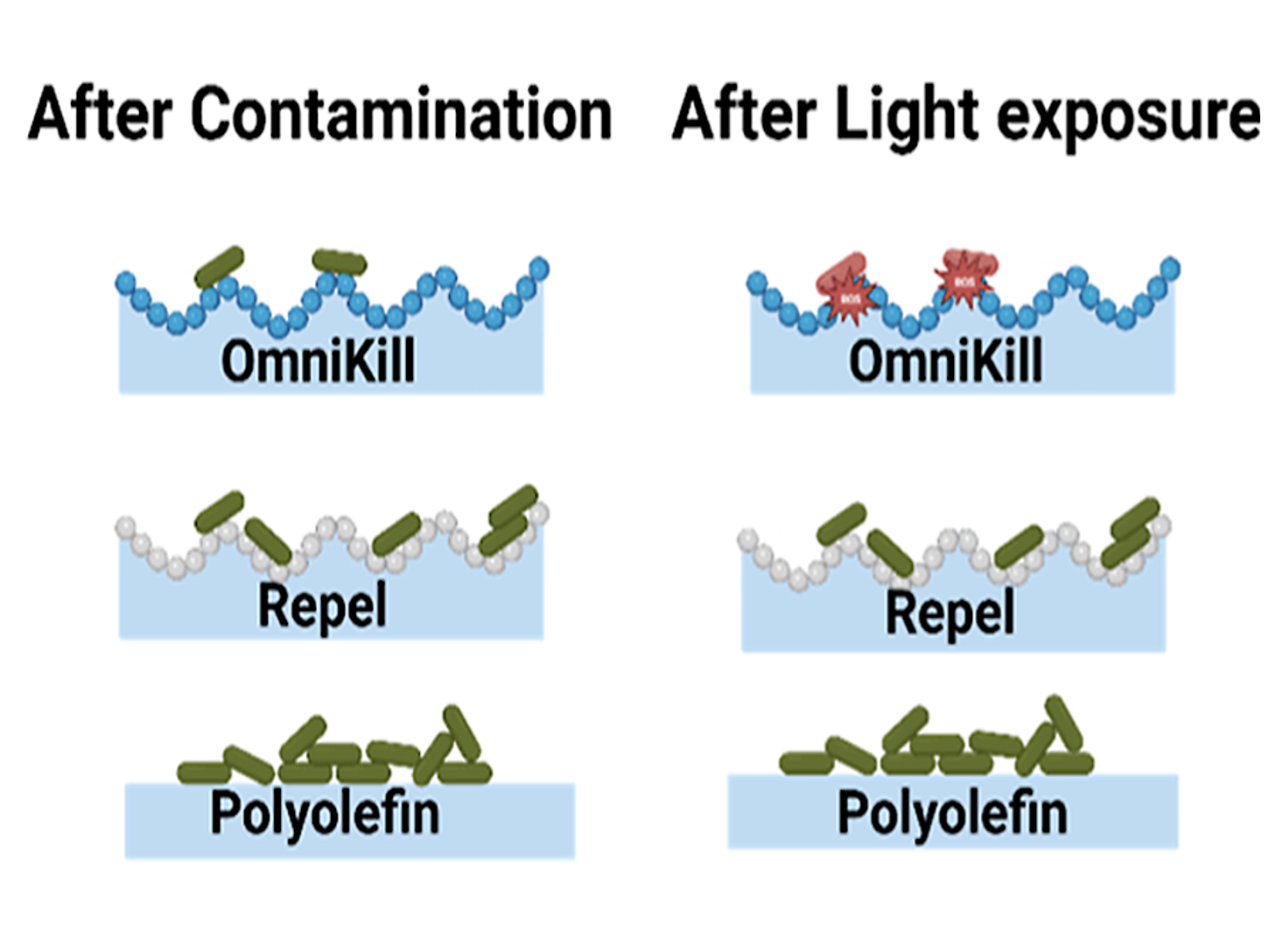
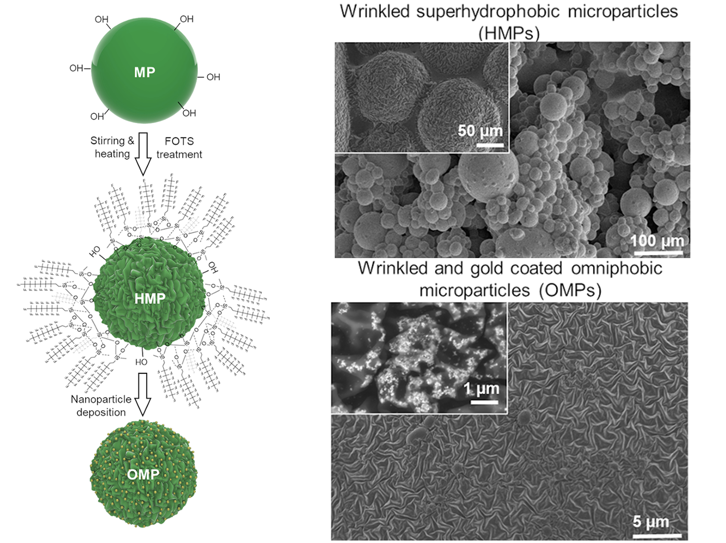
    Superomniphobic and Photoactive Surface Presents Antimicrobial Properties by Repelling and Killing Pathogens

    ACS Applied Materials & Interfaces, 2023

  • Illustration of a membrane-based pathogen detection system. The setup includes food trays at an angle, a fluorescent imaging device, a saturated membrane with a FNAP sensor, and a reagent-saturated membrane. The membrane facilitates pathogen detection by fluorescence, shown with bacteria and a zoomed-in view of bacteria on the membrane.

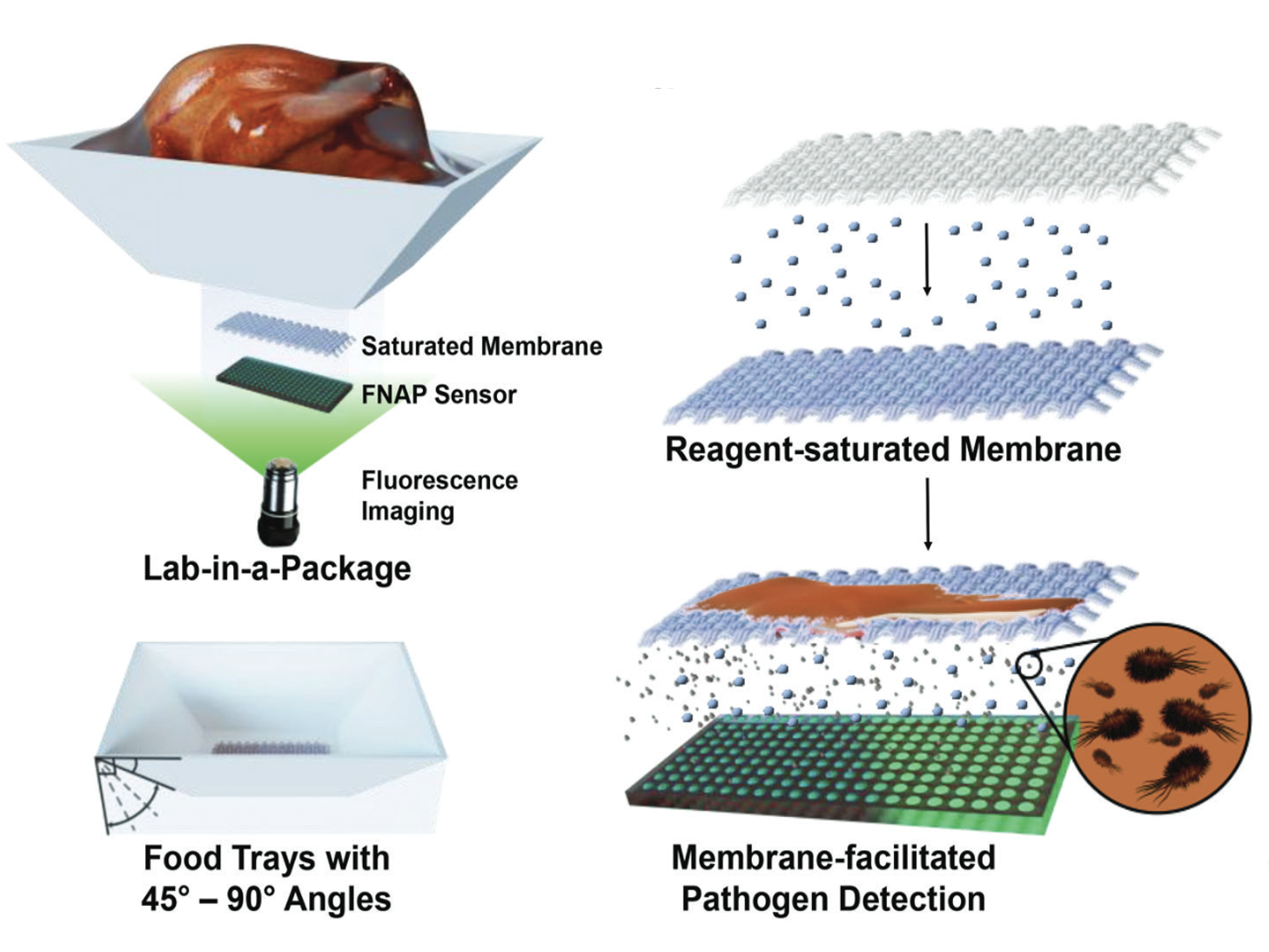
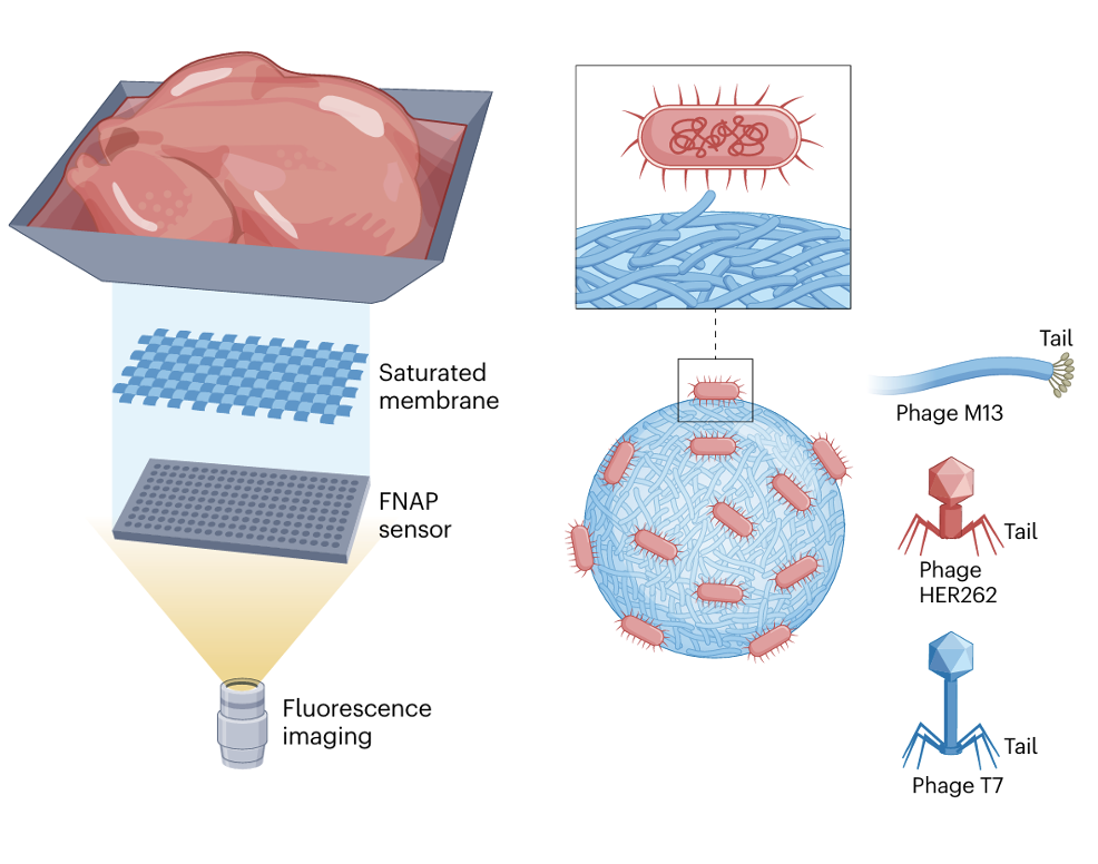
    Advancing in Situ Food Monitoring Through A Smart Lab‐in‐a‐Package System Demonstrated by The Detection of Salmonella in Chicken

    Advanced Materials, 2023

  • Diagram of a DNA biosensor process involving ST spiked food, incubation, filtering, and applying to a tip sensor with Au-tip and nylon. Illustrates DNA hybridization, cleavage, and assembly of Au-on-Au tip sensor components with images of the sensor parts and adsorption pad.

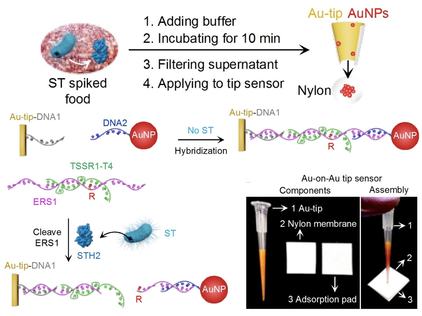
    A Simple Colorimetric Au‐on‐Au Tip Sensor with a New Functional Nucleic Acid Probe for Food‐borne Pathogen Salmonella typhimurium

    Angewandte Chemie, 2023

  • Diagram showing alternative material forms for immune system core recognition agents. It includes sections for pathogens like bacteria and viruses, biogenic amines, and various molecules such as particles, structured materials, anthocyanins, MOFs, oligonucleotides, and antibodies. The diagram also depicts elements like parts of food and bacteria, with labels for pH and various molecular structures.

    Material Breakthroughs in Smart Food Monitoring: Intelligent Packaging and On‐Site Testing Technologies for Spoilage and Contamination Detection

    Advanced Materials, 2023

  • Diagram showing the synthesis process of nanomaterials, starting with microparticles (MP), processed with stirring and FOTS treatment to produce hydroxylated microparticles (HMP), which are further converted into nanoparticle-decorated microparticles (OHP). The right side displays electron microscope images of wrinkled superhydrophobic microparticles (HMPs), wrinkled and gold-coated omniphobic microparticles (OMPs), with scales of 50, 100, 1, and 5 micrometers.

    A Bifunctional Spray Coating Reduces Contamination on Surfaces by Repelling and Killing Pathogens

    ACS Applied Materials & Interfaces, 2023

  • Diagram showing the process of transforming PDMS into functionalized gold nanoparticles for biomedical applications through chemical treatment, heating, and surface modification, accompanied by electron microscope images of different nanostructures.

    An Omniphobic Spray Coating Created from Hierarchical Structures Prevents the Contamination of High‐Touch Surfaces with Pathogens

    Small, 2023

  • Microscopic images and schematic diagrams showing the structure, synthesis, and crosslinking of phage microgels and aerogel microparticles, including electron micrographs and labeled illustrations of different microgel types and their crosslinking methods.

    Self-assembling Nanofibrous Bacteriophage Microgels as Sprayable Antimicrobials Targeting Multidrug-resistant Bacteria

    Nature Communications, 2022

  • A scientific process diagram showing four stages: extrusion of polymer blends, hot pressing into transparent PVDF/PMMA film, swelling and coagulation of a surface to create a hierarchically structured surface, and lubricant infusion resulting in a transparent slippery liquid-infused surface (LIS).

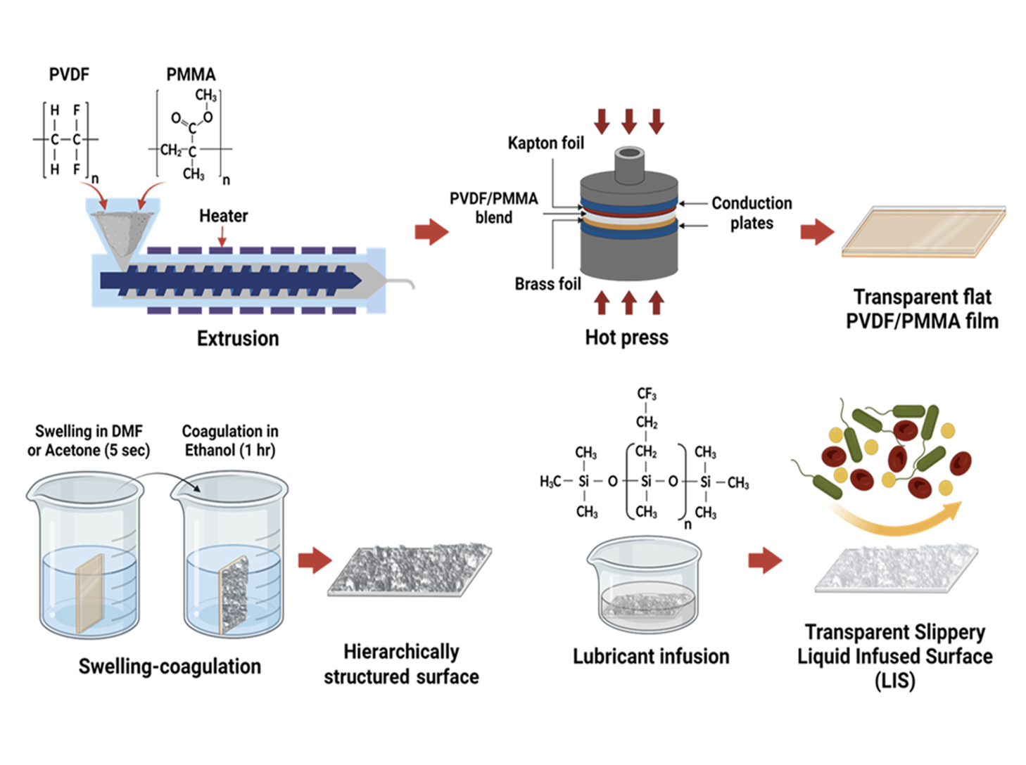
    Highly Stable Hierarchically Structured All-Polymeric Lubricant-Infused Films Prevent Thrombosis and Repel Multidrug-Resistant Pathogens

    ACS Applied Materials & Interfaces, 2022

  • Diagram showing a biosensor with a purple detection surface, three white tubes connected to a purple surface, and illustrations of molecular binding and electron transfer processes.

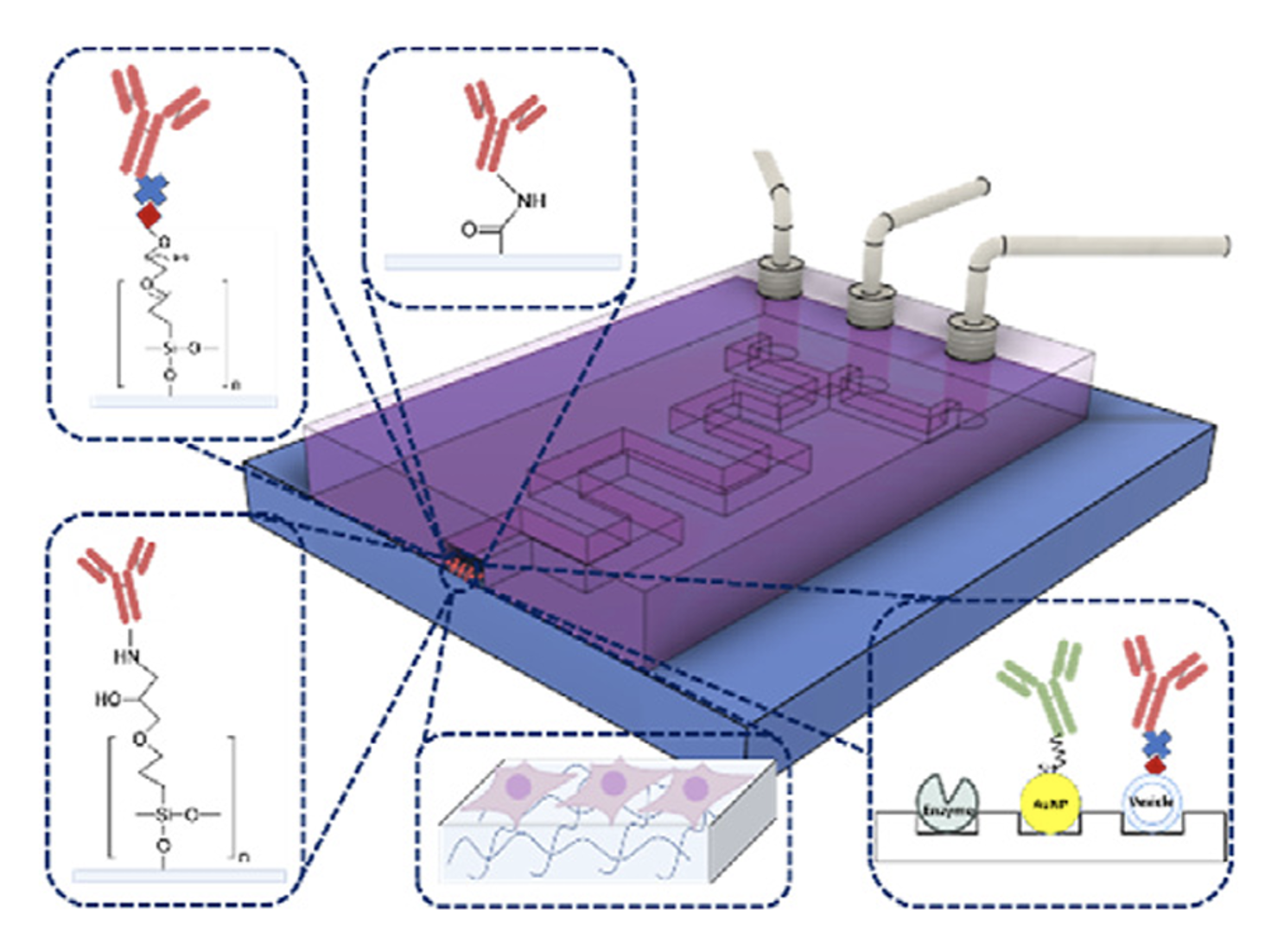
    Bio-functionalization of Microfluidic Platforms Made of Thermoplastic Materials: A Review

    Analytica Chimica Acta, 2022

  • Diagram showing bacterial repellency and antithrombotic properties of planar and hierarchical PDMS. Top row displays bacteria and bacteria-resistant images with fluorescent microscopy. Bottom row shows antithrombotic images via fluorescence microscopy for both PDMS types, with grayscale scale bar of 50 micrometers.

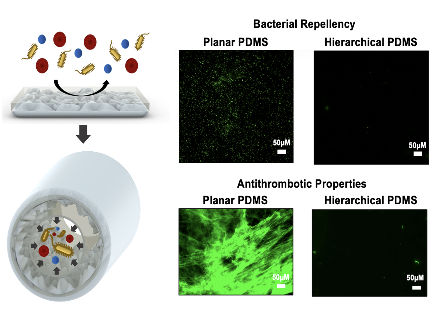
    Transparent and Highly Flexible Hierarchically Structured Polydimethylsiloxane Surfaces Suppress Bacterial Attachment and Thrombosis Under Static and Dynamic Conditions

    Small, 2022

  • Microscopic images comparing a hierarchical surface with a polysiloxane nanostructure coating. Left image shows a rough surface at 100 micrometers scale, right image shows a nanostructured surface at 4 micrometers scale.

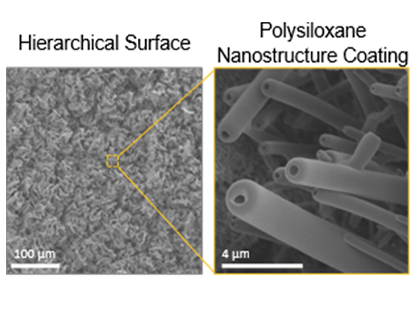
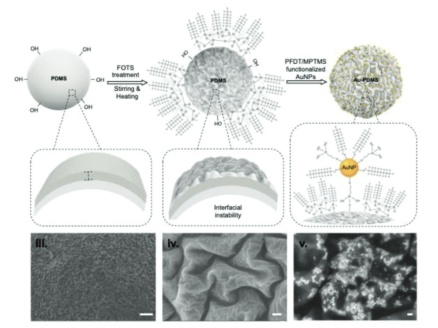
    Producing Fluorine-and Lubricant-Free Flexible Pathogen-and Blood-Repellent Surfaces Using Polysiloxane-Based Hierarchical Structures

    ACS Applied Materials & Interfaces, 2022

  • Diagram illustrating the process of manufacturing and bonding using laser technology and various methods. The left side shows fabrication techniques like hot embossing, injection molding, laser ablation, micro-milling, and 3D printing. The right side lists bonding techniques such as thermal fusion, solvent bonding, chemical bonding, welding, and adhesive layers.

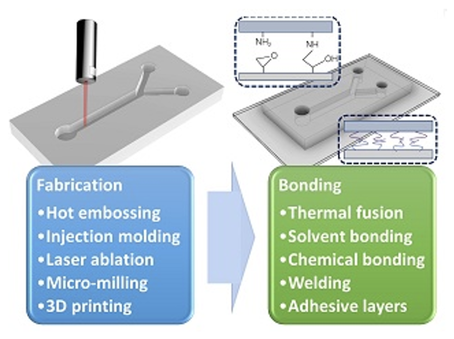
    The Fabrication and Bonding of Thermoplastic Microfluidics: A Review

    Materials, 2022

  • Comparison of traditional biosensors and LISYME biosensors. The traditional biosensor shows biofouling and unwanted surface adhesion on the sensor surface. The LISYME biosensor illustrates less biofouling and unwanted surface adhesion, indicating improved resistance to fouling and enhanced sensor performance.

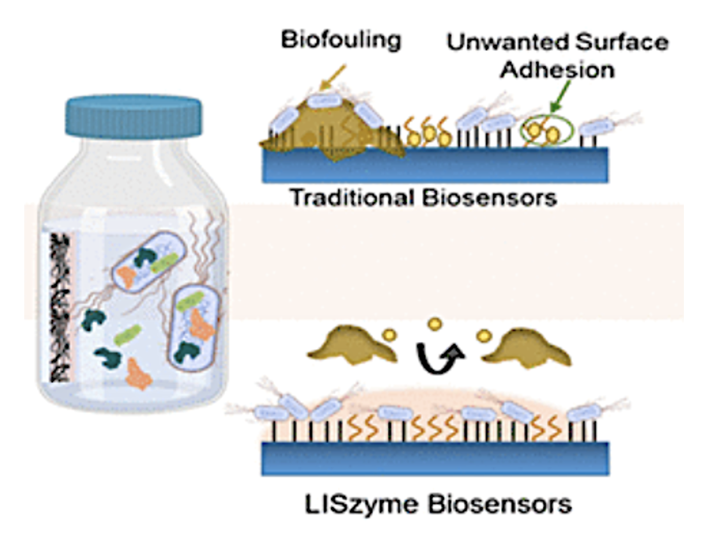
    LISzyme Biosensors: DNAzymes Embedded in an Anti-biofouling Platform for Hands-free Real-time Detection of Contamination in Milk

    ACS Nano, 2021

  • Diagram showing strategies for biosensing and immobilization involving DNA, proteins, and nanoparticles with colored illustrations of bacteria, metals, and molecules.

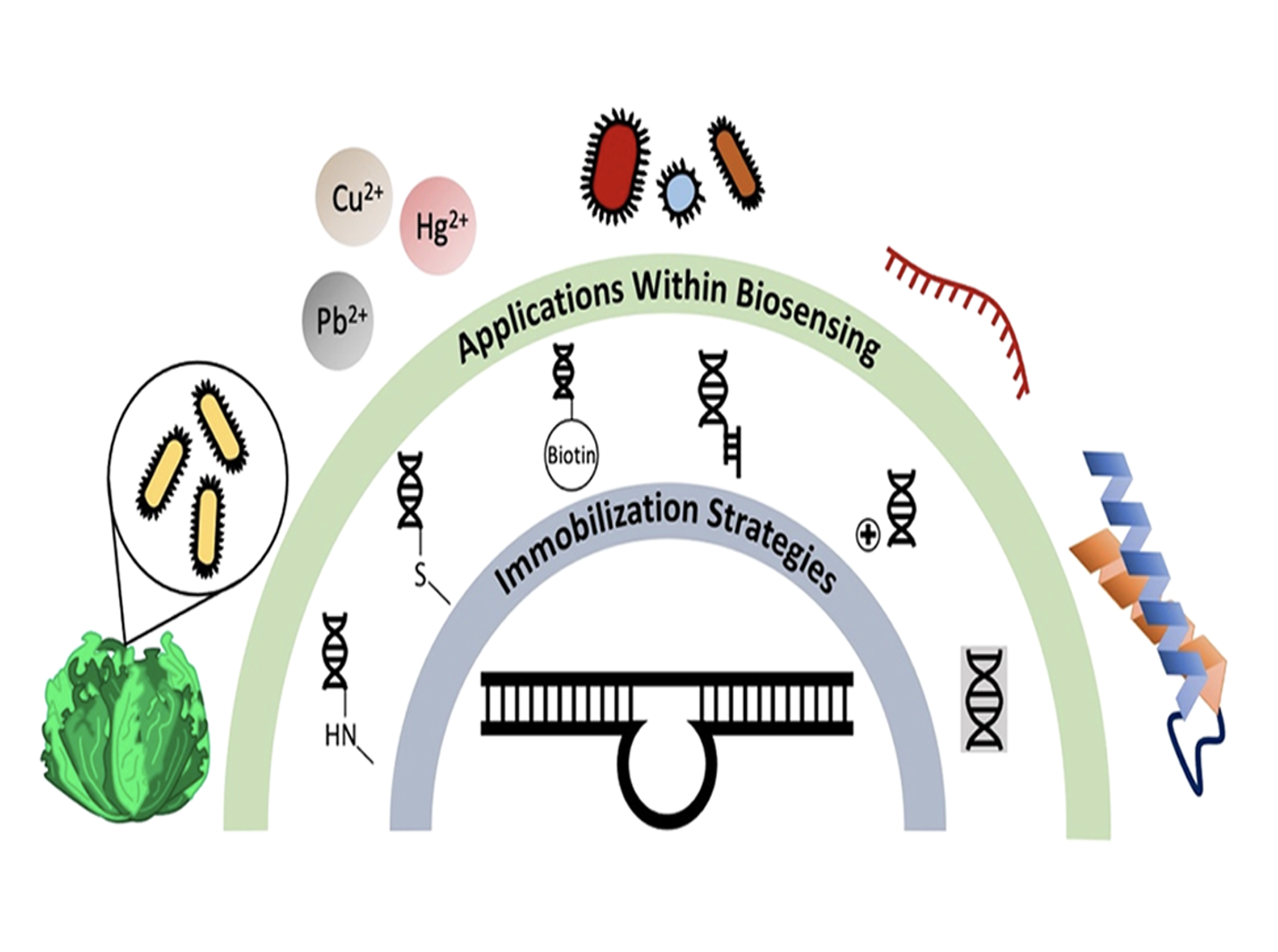
    DNAzyme-Based Biosensors: Immobilization Strategies, Applications, and Future Prospective

    ACS Nano, 2021

  • Diagram showing biomedical device fabrication processes, including hydrolyzation, chemical bonding, adhesive films, antibody, cells, other proteins, with labels for each process and visualization of molecular structures.

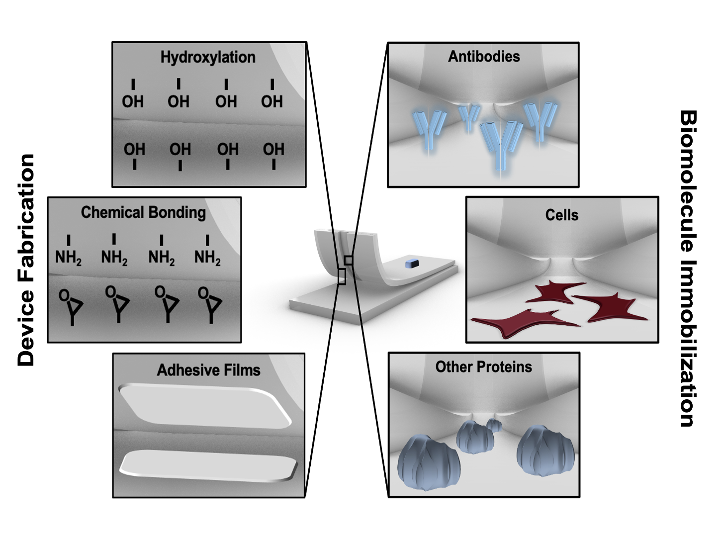
    Conventional and Emerging Strategies for the Fabrication and Functionalization of PDMS-based Microfluidic Devices

    Lab on a Chip, 2021

  • Scientific diagram showing experimental steps, microscopy images, and a graph related to cell imaging and analysis with different buffers, stainings, and microscopy techniques labeled as panels a through i.

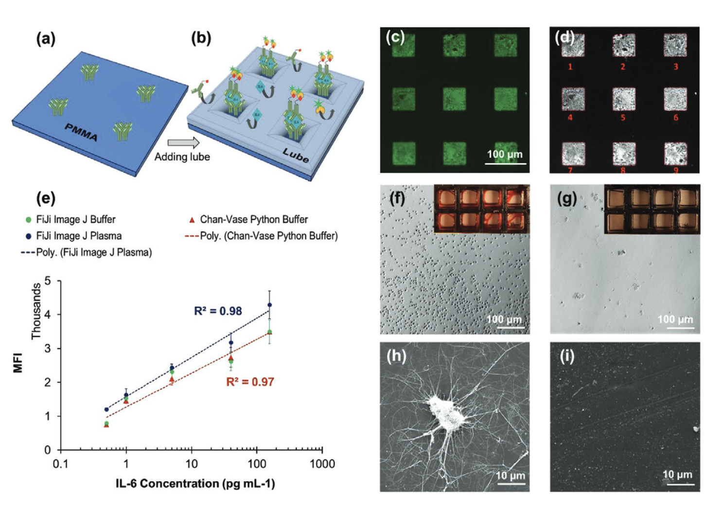
    Antibody Micropatterned Lubricant‐Infused Biosensors Enable Sub‐Picogram Immunofluorescence Detection of Interleukin 6 in Human Whole Plasma

    Small, 2020